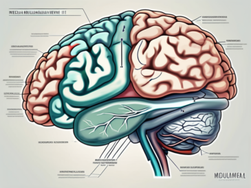
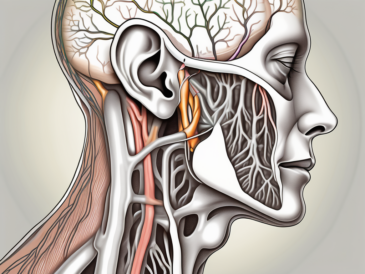
The glossopharyngeal nerve, a vital component of the human nervous system, plays a crucial role in the transfer of sensory and motor information. To fully comprehend the significance of this nerve, it is essential to explore its anatomical structure, functions, and its connection to the brain. Furthermore, it is pertinent to understand the implications that damage to the glossopharyngeal nerve can have on an individual’s well-being. Advancements in neurological research have shed light on the intricate workings of this nerve, which may pave the way for new treatment and therapy options.
Understanding the Glossopharyngeal Nerve
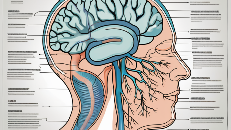
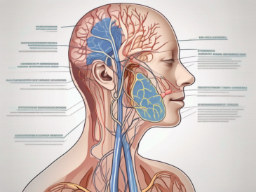
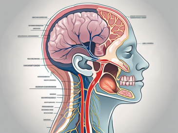
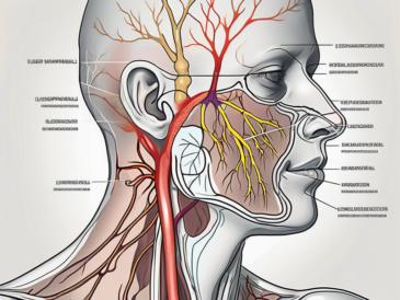
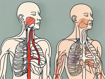
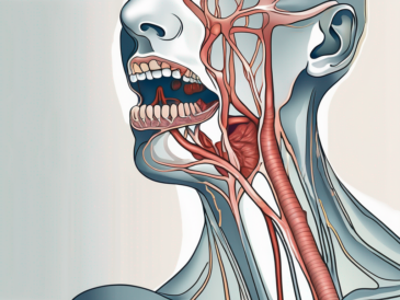
The glossopharyngeal nerve, also known as cranial nerve IX, is one of the twelve cranial nerves originating from the brain. It emerges from the medulla oblongata, a vital part of the brainstem, and extends down into the back of the throat, neck, and middle ear region. Branches of the glossopharyngeal nerve innervate various structures, including the tongue, pharynx, tonsils, and salivary glands.
The anatomy of the glossopharyngeal nerve is fascinating. It is composed of both sensory and motor fibers, allowing it to perform a wide range of functions. The sensory fibers carry information from the tongue, pharynx, tonsils, and middle ear, providing us with crucial sensations. For example, when we taste food, the sensory fibers of the glossopharyngeal nerve transmit the signals to our brain, allowing us to experience different flavors. Additionally, these fibers also play a role in monitoring blood pressure and oxygen levels in the carotid arteries, ensuring our body functions optimally.
On the other hand, the motor fibers of the glossopharyngeal nerve are responsible for controlling certain muscles involved in swallowing and initiating the gag reflex. When we swallow, these motor fibers coordinate the movement of our throat muscles, allowing food and liquids to pass through smoothly. Similarly, when something triggers the gag reflex, the motor fibers of the glossopharyngeal nerve cause the muscles in the back of our throat to contract, protecting our airway from potential harm.
Understanding the functions of the glossopharyngeal nerve is crucial in comprehending the complexity of our nervous system. This nerve not only enables us to taste and swallow but also plays a role in regulating our cardiovascular system. Its intricate network of sensory and motor fibers highlights the remarkable nature of our body’s design.
The Brain’s Reception of Information
The brain is an incredibly complex organ that plays a vital role in receiving and processing sensory information. When it comes to the glossopharyngeal nerve, it is crucial to understand how the brain receives and interprets the signals it sends.
The Role of the Brain’s Lobes
The human brain is divided into different regions called lobes, each with its own unique functions. One lobe, in particular, plays a significant role in processing sensory information related to taste, which is directly relevant to the glossopharyngeal nerve.
The parietal lobe, located towards the top and back of the brain, is responsible for integrating sensory information from various parts of the body. It plays a crucial role in processing taste sensations received from the glossopharyngeal nerve.
Within the parietal lobe, there is a specific region known as the primary gustatory cortex. This area is dedicated to processing taste information and is where the brain differentiates between various tastes, such as sweet, sour, salty, and bitter, based on the signals received from the glossopharyngeal nerve.
Information Processing in the Brain
Upon receiving sensory input from the glossopharyngeal nerve, the brain begins a complex process of information processing. The sensory signals from the tongue and other relevant structures are relayed to the primary gustatory cortex within the parietal lobe.
Once the signals reach the primary gustatory cortex, the brain starts to analyze and interpret the taste information. This analysis involves comparing the incoming signals with stored information about different tastes, acquired through previous experiences.
As the brain processes the taste information, it also integrates it with other sensory inputs, such as smell and texture, to create a complete perception of taste. This integration allows us to fully experience and appreciate the flavors of the food we consume.
Furthermore, the brain’s processing of taste information goes beyond simple recognition. It also plays a role in determining our preferences and aversions to certain tastes. These preferences can be influenced by a variety of factors, including genetics, cultural influences, and individual experiences.
Overall, the brain’s reception and processing of sensory information, including taste signals from the glossopharyngeal nerve, is a complex and fascinating process. It involves the integration of signals from various parts of the body and the interpretation of these signals to create our perception of taste.
The Glossopharyngeal Nerve and the Brain Connection
Pathway of the Glossopharyngeal Nerve
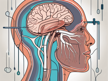
To understand which specific lobe receives information from the glossopharyngeal nerve, it is crucial to explore the pathway this nerve takes within the brain. After leaving the medulla oblongata, the glossopharyngeal nerve enters the skull through the jugular foramen, a narrow opening located between the temporal bone and the occipital bone. This intricate journey through the skull allows the nerve to reach its destination and fulfill its vital functions.
Once inside the skull, the glossopharyngeal nerve branches out into smaller nerve fibers, resembling a complex network of communication pathways. These fibers spread throughout the brain, forming connections with different regions and structures. By doing so, the glossopharyngeal nerve ensures that the sensory information it carries can be efficiently transmitted and processed.
These smaller nerve fibers transmit sensory information to various regions of the brain, including the parietal lobe. The parietal lobe, located at the upper back area of the brain, plays a crucial role in processing sensory input from different parts of the body. It is responsible for integrating and interpreting sensory information, allowing individuals to perceive and make sense of their surroundings.
The Specific Lobe Receiving Information
As mentioned earlier, the parietal lobe is primarily responsible for processing sensory input related to taste. It interprets the signals received from the glossopharyngeal nerve, allowing individuals to experience various tastes and differentiate between them. This remarkable ability to perceive taste is made possible by the intricate connection between the glossopharyngeal nerve and the parietal lobe.
Within the parietal lobe, there are specific areas known as gustatory cortex, which are dedicated to processing taste information. These areas receive signals from the glossopharyngeal nerve and work in harmony with other brain regions to create the rich and complex experience of taste. The gustatory cortex not only helps identify different tastes but also contributes to the overall enjoyment and appreciation of food and beverages.
Furthermore, the parietal lobe’s involvement in taste perception further emphasizes the importance of the glossopharyngeal nerve’s connection to this specific region of the brain. Without this connection, the ability to taste and savor the flavors of our favorite foods would be greatly diminished.
Implications of Damage to the Glossopharyngeal Nerve
The glossopharyngeal nerve plays a crucial role in the functioning of the throat and mouth. Damage or dysfunction of this nerve can have severe implications for an individual’s overall health and well-being. It is important to understand the symptoms, treatment options, and recovery process associated with glossopharyngeal nerve damage.
Symptoms of Glossopharyngeal Nerve Damage
When the glossopharyngeal nerve is damaged, various symptoms may arise, affecting different aspects of a person’s daily life. One common symptom is difficulty swallowing, also known as dysphagia. This can make it challenging to eat and drink, leading to weight loss and malnutrition if not properly managed.
Another symptom of glossopharyngeal nerve damage is changes in taste perception. The taste buds located on the back of the tongue are innervated by this nerve, so any dysfunction can result in a loss or alteration of taste. This can greatly impact a person’s enjoyment of food and beverages, as well as their ability to detect potential dangers, such as spoiled or toxic substances.
Throat pain is also a common symptom of glossopharyngeal nerve damage. The nerve supplies sensation to the back of the throat, so any injury or dysfunction can lead to persistent discomfort or a sore throat. This can make speaking, swallowing, and even breathing painful and challenging.
In some cases, glossopharyngeal nerve damage can cause the presence of a persistently acute gag reflex. This means that even minor stimuli, such as brushing the back of the throat with a toothbrush, can trigger a strong gag reflex. This can be distressing and may further contribute to difficulties with eating, drinking, and maintaining oral hygiene.
Treatment and Recovery Options
If an individual experiences symptoms suggestive of glossopharyngeal nerve damage, it is vital to seek medical attention promptly. A healthcare professional will perform a thorough clinical evaluation to assess the extent of the nerve dysfunction. This evaluation may include imaging studies, nerve conduction tests, and other specialized investigations to determine the underlying cause of the damage.
Treatment options for glossopharyngeal nerve damage depend on the specific circumstances and underlying cause. In some cases, medication may be prescribed to manage pain, reduce inflammation, or address any underlying conditions contributing to the nerve dysfunction. Physical therapy may also be recommended to improve swallowing function and strengthen the muscles involved in speech and swallowing.
In severe cases, surgical intervention may be necessary. This could involve repairing the damaged nerve or removing any structures compressing or impinging on the nerve. Surgical procedures are typically considered when conservative treatments have failed to provide relief or when there is a significant risk of further complications.
Recovery from glossopharyngeal nerve damage can vary depending on the extent of the injury and the individual’s overall health. It is crucial for individuals to consult with a doctor who can provide appropriate guidance and develop a tailored treatment plan. Rehabilitation and ongoing support may be necessary to help individuals regain their ability to eat, speak, and enjoy the sense of taste.
In conclusion, glossopharyngeal nerve damage can have significant implications for an individual’s quality of life. It is important to recognize the symptoms, seek timely medical attention, and explore appropriate treatment options to optimize recovery and minimize the impact of this condition.
The Future of Neurological Research
Advancements in Understanding the Glossopharyngeal Nerve
Neurological research continues to expand our understanding of the intricate mechanisms governing the glossopharyngeal nerve. Scientists are working diligently to uncover the finer details of how this nerve functions, and how its damage can be prevented or mitigated. Exploring the precise neural pathways involved in taste perception facilitated by the glossopharyngeal nerve may pave the way for exciting advancements in the field of sensory research.
The glossopharyngeal nerve, also known as the ninth cranial nerve, is a complex structure that plays a crucial role in transmitting sensory information from the tongue, throat, and other regions of the oral cavity to the brain. It is responsible for carrying taste signals from the posterior third of the tongue, as well as providing sensory innervation to the tonsils, pharynx, and middle ear.
Recent studies have focused on unraveling the intricate connections between the glossopharyngeal nerve and the brain. By utilizing advanced imaging techniques, researchers have been able to map the neural pathways associated with taste perception. This mapping process involves tracing the nerve fibers from the tongue to the brain, allowing scientists to identify specific regions that are activated during taste sensation.
Furthermore, investigations into the glossopharyngeal nerve have revealed its close relationship with other cranial nerves involved in the sensory processing of the oral cavity. The trigeminal nerve, for instance, works in tandem with the glossopharyngeal nerve to provide a comprehensive sensory experience. Understanding the interplay between these nerves is crucial for developing effective treatments for taste disorders and other related conditions.
Potential Impact on Treatment and Therapy
As our knowledge of the glossopharyngeal nerve deepens, there is a growing potential for groundbreaking advancements in treatment and therapy options. Utilizing a comprehensive understanding of the glossopharyngeal nerve’s role in taste perception, researchers may devise targeted therapies that could enhance or restore gustatory functions in individuals with damaged or dysfunctional nerves. However, it is important to note that further research and clinical trials are necessary to validate these prospects and translate them into effective therapeutic interventions.
One potential avenue for therapeutic intervention lies in the field of regenerative medicine. Scientists are exploring the possibility of using stem cells to regenerate damaged glossopharyngeal nerve fibers, thereby restoring normal sensory function. This approach holds great promise for individuals suffering from taste disorders caused by nerve damage or degeneration.
Another area of interest is the development of neuroprosthetic devices that can bypass damaged glossopharyngeal nerves and directly stimulate the brain’s taste centers. These devices, often referred to as brain-computer interfaces, have shown promising results in preliminary studies. By electrically stimulating the appropriate regions of the brain, these interfaces could potentially restore taste perception in individuals with severe nerve damage.
In addition to treatment options, a deeper understanding of the glossopharyngeal nerve could also lead to advancements in diagnostic techniques. Currently, the diagnosis of glossopharyngeal nerve dysfunction relies on a combination of clinical evaluation and specialized tests. However, with further research, it may be possible to develop more accurate and efficient diagnostic tools, allowing for earlier detection and intervention.
Furthermore, the implications of glossopharyngeal nerve research extend beyond taste perception. This nerve also plays a role in regulating blood pressure and heart rate, making it a potential target for the treatment of cardiovascular disorders. By unraveling the complexities of the glossopharyngeal nerve’s involvement in these physiological processes, researchers may uncover new therapeutic strategies for managing conditions such as hypertension and arrhythmias.
In conclusion, the glossopharyngeal nerve, while often overshadowed by its counterparts in discussions of the nervous system, plays a vital role in taste perception and other essential sensory functions. As research in the field of neuroscience progresses, our understanding of the intricacies of this nerve and its connection to the brain continues to expand. By elucidating the anatomy, functions, and implications of damage to the glossopharyngeal nerve, healthcare professionals can better diagnose and manage conditions affecting this critical neural pathway. Ultimately, ongoing advancements in neurological research hold promising implications for enhancing treatment options and improving the quality of life for individuals experiencing glossopharyngeal nerve dysfunction.