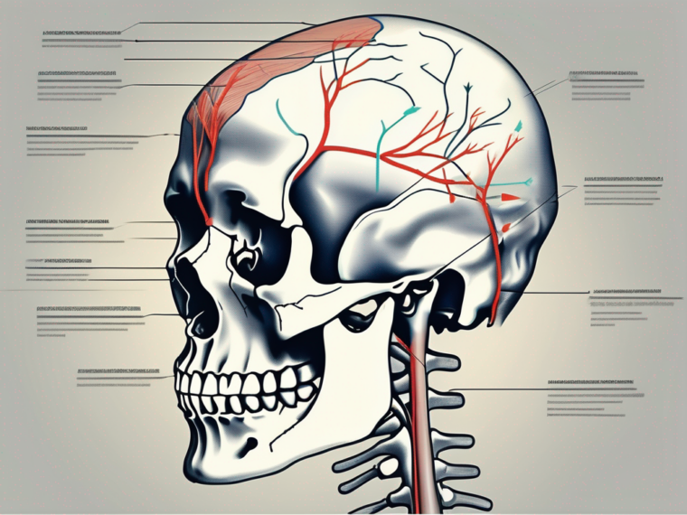
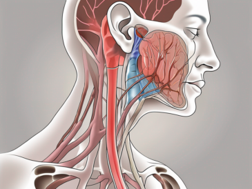
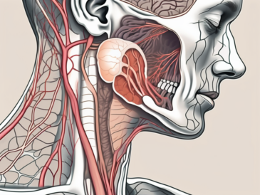
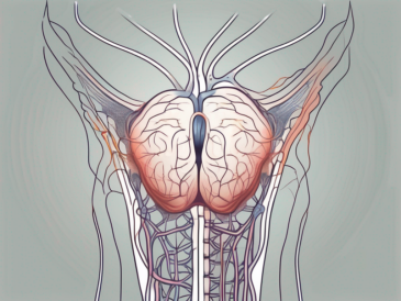
The glossopharyngeal nerve is one of the twelve cranial nerves that emerge from the brainstem. It plays a crucial role in the innervation of various structures within the head and neck region. Understanding the pathway of axons through the foramen is essential in comprehending the functions and implications of this nerve.
Understanding the Glossopharyngeal Nerve
The glossopharyngeal nerve, also known as cranial nerve IX, is responsible for sensory and motor functions in the oral cavity and throat. Its name originates from the Greek words “glossa” meaning tongue and “pharynx” referring to the throat. This nerve carries sensory information from the posterior third of the tongue, the pharynx, the middle ear, and part of the palate. Additionally, it supplies motor innervation to certain muscles involved in swallowing and speech production.
Anatomy of the Glossopharyngeal Nerve
The glossopharyngeal nerve emerges from the medulla oblongata, a region located at the base of the brainstem. It originates between the olive and the inferior cerebellar peduncle. Cranial nerve IX consists of both sensory and motor fibers, which travel together until they reach a structure called the jugular foramen.
As the glossopharyngeal nerve travels through the jugular foramen, it branches out into different directions, providing innervation to various structures. One branch extends towards the posterior third of the tongue, where it plays a crucial role in transmitting taste sensations. Another branch extends towards the pharynx, relaying sensory information from this region, including the tonsils. Additionally, the glossopharyngeal nerve sends branches towards the middle ear, contributing to the perception of sound and maintaining balance.
Within the medulla oblongata, the glossopharyngeal nerve is surrounded by other cranial nerves, forming a complex network of neural connections. This intricate arrangement allows for the integration of sensory and motor signals, enabling precise control over the functions of the oral cavity and throat.
Function of the Glossopharyngeal Nerve
The glossopharyngeal nerve serves several crucial functions within the body. Sensory functions include transmitting taste sensations from the posterior third of the tongue, assisting in the regulation of blood pressure and heart rate through sensory input from the carotid sinus, and providing general sensation to the pharynx.
In addition to its sensory functions, the glossopharyngeal nerve plays a vital role in motor functions related to swallowing. The motor fibers of this nerve innervate the stylopharyngeus muscle, a key player in the swallowing process. When food or liquid is ingested, the stylopharyngeus muscle contracts, elevating the larynx and opening the upper esophageal sphincter. This coordinated movement prevents aspiration of food or liquids into the airway, ensuring proper nutrition and respiration.
Furthermore, the glossopharyngeal nerve contributes to the reflexive response of the gag reflex. When the back of the throat is stimulated, such as by touching the tonsils, the sensory fibers of the glossopharyngeal nerve transmit this information to the brainstem, triggering a protective reflex that helps prevent choking or the entry of foreign objects into the airway.
Overall, the glossopharyngeal nerve plays a crucial role in the complex functions of the oral cavity and throat. Its sensory and motor functions ensure the proper functioning of taste perception, blood pressure regulation, swallowing, and the protection of the airway. Understanding the anatomy and function of this nerve is essential for comprehending the intricate mechanisms involved in these vital processes.
The Journey of Motor Neurons
Motor neurons play a crucial role in transmitting signals from the brain to muscles, allowing voluntary movements and controlling various bodily functions. In the case of the glossopharyngeal nerve, motor neurons are vital for the proper functioning of the muscles involved in swallowing and speech production.
The Role of Motor Neurons in the Glossopharyngeal Nerve
The motor neurons innervating the glossopharyngeal nerve have a specific role in coordinating the complex process of swallowing. These neurons provide the necessary muscle contractions to propel food and liquids from the mouth through the pharynx and into the esophagus. They also contribute to the elevation of the larynx, a protective mechanism preventing choking and aspiration.
Swallowing is a remarkable process that involves a series of intricate movements orchestrated by motor neurons. When we consume food or drink, the muscles in our mouth and throat work together to form a cohesive action. The motor neurons within the glossopharyngeal nerve are responsible for coordinating these movements, ensuring that the food or liquid is safely transported from the mouth to the esophagus.
Imagine a delicious meal in front of you. As you take a bite and begin to chew, the motor neurons in the glossopharyngeal nerve send signals to the muscles in your mouth, instructing them to break down the food into smaller, more manageable pieces. These neurons then coordinate the contraction of the muscles in your throat, propelling the chewed food towards the back of your mouth.
Once the food reaches the back of your mouth, the motor neurons continue their work. They activate the muscles in your pharynx, causing a series of coordinated contractions that push the food further down the throat. This process, known as peristalsis, ensures that the food is propelled in a controlled manner, preventing any blockages or discomfort.
As the food enters the esophagus, the motor neurons within the glossopharyngeal nerve play a crucial role in initiating the next phase of swallowing. They coordinate the relaxation of the muscles in the upper esophagus, allowing the food to pass through and enter the stomach. Without these motor neurons, the process of swallowing would be disrupted, leading to difficulties in eating and potential health complications.
Pathway of Motor Neurons
Motor neurons of the glossopharyngeal nerve travel within the nerve as it courses through the skull and neck. They join the sensory fibers of the glossopharyngeal nerve and travel together until they reach the jugular foramen, a crucial landmark in their journey.
The journey of motor neurons within the glossopharyngeal nerve is a fascinating one. Starting from the brain, these neurons extend their long, slender projections called axons, which travel through the protective bony structures of the skull and neck. Along the way, they navigate through complex networks of blood vessels, connective tissues, and other nerves, ensuring that they reach their intended destination.
As the motor neurons travel within the glossopharyngeal nerve, they interact with the sensory fibers of the nerve. This interaction allows for the seamless coordination between sensory and motor functions, ensuring that the information gathered by the sensory fibers is translated into appropriate motor responses.
Upon reaching the jugular foramen, the motor neurons continue their journey, branching out to innervate the specific muscles involved in swallowing and speech production. These branches extend like intricate pathways, connecting with the target muscles and forming the necessary neural connections for proper functioning.
It is truly remarkable how motor neurons navigate through the intricate pathways of the glossopharyngeal nerve, ensuring that the muscles involved in swallowing and speech production receive the necessary signals for coordinated movement. Without these motor neurons, the complex process of swallowing would be disrupted, leading to difficulties in eating and communicating effectively.
The Foramen in Focus
The foramen is a term used in anatomy to describe an opening or passage through which nerves, blood vessels, or other structures pass. In the case of the glossopharyngeal nerve, the foramen plays a significant role in the transit of motor neurons.
The glossopharyngeal nerve, also known as cranial nerve IX, is one of the twelve cranial nerves that emerge directly from the brain. It is responsible for providing motor and sensory innervation to the tongue, throat, and certain glands in the head and neck region. The foramen, in this context, acts as a gateway for the glossopharyngeal nerve to reach its target tissues and carry out its vital functions.
Defining the Foramen
The glossopharyngeal nerve, along with cranial nerve X (the vagus nerve) and cranial nerve XI (the accessory nerve), all pass through a structure known as the jugular foramen. Situated at the base of the skull, the jugular foramen serves as a conduit for several important structures.
Within the jugular foramen, the glossopharyngeal nerve intertwines with the vagus nerve and the accessory nerve, forming a complex network of neural connections. This convergence allows for efficient communication and coordination between these cranial nerves, ensuring the proper functioning of various physiological processes.
Furthermore, the jugular foramen is not solely dedicated to the passage of nerves. It also accommodates the internal jugular vein, a major blood vessel responsible for draining deoxygenated blood from the brain and face. The coexistence of nerves and blood vessels within the jugular foramen highlights the intricate relationship between the nervous and circulatory systems.
During their transit through the jugular foramen, the motor neurons of the glossopharyngeal nerve meet with sensory fibers originating from other regions. This union allows for their coordinated functioning and integration of sensory and motor responses.
Different Types of Foramen and Their Functions
Various foramina exist within the skull, each accommodating specific structures. The jugular foramen, in particular, facilitates the passage of not only the glossopharyngeal nerve but also the vagus and accessory nerves. These nerves are essential for coordinating various functions such as swallowing, vocalization, and the movement of the head and neck.
Another noteworthy foramen is the foramen magnum, which is located at the base of the skull and serves as the entry point for the spinal cord. This crucial opening allows for the connection between the brain and the rest of the body, enabling the transmission of signals and the coordination of voluntary and involuntary movements.
Understanding the types and functions of different foramina is imperative for comprehending the intricate network of nerves and blood vessels within the skull and their respective roles in maintaining proper bodily function. The complex interplay between these structures ensures the seamless integration of sensory and motor information, allowing us to perform essential activities such as eating, speaking, and moving with precision and efficiency.
The Passage of Axons through the Foramen
The axons of motor neurons within the glossopharyngeal nerve must navigate through the jugular foramen to reach their target muscles. This process ensures the precise coordination of swallowing and other essential functions.
The Process of Axon Passage
As motor neurons, originating in the medulla oblongata, travel within the glossopharyngeal nerve, they approach the jugular foramen. This narrow opening requires these axons to navigate through tight spaces while avoiding compression or damage.
The intricate anatomy of the jugular foramen prevents overcrowding and potential entrapment of axons, ensuring the smooth passage of motor neurons required for proper function.
Within the jugular foramen, the axons of motor neurons undergo a series of remarkable adaptations to facilitate their passage. These adaptations include changes in the axonal membrane, allowing for increased flexibility and resistance to compression. Additionally, specialized glial cells within the foramen provide structural support and guidance, ensuring the axons stay on the correct path.
Furthermore, the jugular foramen is lined with a thin layer of protective connective tissue known as the meninges. This layer acts as a cushion, shielding the axons from potential external pressures or trauma. The meninges also play a crucial role in maintaining the optimal environment for axonal function, providing necessary nutrients and removing waste products.
The Importance of the Foramen for Axons
The jugular foramen acts as a conduit, allowing for the precise passage of axons, including those of motor neurons, within the glossopharyngeal nerve. This structural feature safeguards against compression, entrapment, or damage to these critical components of the neural circuit.
Any hindrance or blockage within the foramen may impede the proper functioning of these motor neurons, leading to potential complications and impairments in swallowing, speech, and other associated functions.
Moreover, the jugular foramen serves as a vital connection point between the central nervous system and the peripheral nervous system. It not only allows the axons to reach their target muscles but also facilitates the exchange of information between the brain and the periphery. This exchange is crucial for the coordination of various physiological processes, such as the regulation of blood pressure and heart rate.
Interestingly, the size and shape of the jugular foramen can vary among individuals, highlighting the uniqueness of each person’s anatomy. This anatomical variation can have implications for the passage of axons, as individuals with a narrower foramen may experience more challenges in motor neuron navigation.
In conclusion, the jugular foramen plays a pivotal role in ensuring the smooth passage of axons, particularly those of motor neurons within the glossopharyngeal nerve. Its intricate anatomy, coupled with specialized adaptations and protective mechanisms, allows for the precise coordination of essential functions. Understanding the importance of this structural feature provides valuable insights into the complexity of the neural circuitry and the remarkable adaptability of the human body.
Implications of Foramen Blockage or Damage
The blockage or damage of the jugular foramen, through which the axons of motor neurons pass, can have significant consequences for the functioning of the glossopharyngeal nerve and associated muscles.
When the jugular foramen is blocked or damaged, it can disrupt the normal flow of neural signals, leading to various complications. These complications can range from mild discomfort to severe impairment in the affected individual’s ability to perform everyday tasks.
One potential consequence of foramen blockage or damage is the development of swallowing difficulties. The glossopharyngeal nerve plays a crucial role in coordinating the muscles involved in swallowing. When the neural signals cannot pass through the jugular foramen properly, it can result in a disruption of this coordination, leading to problems with swallowing food and liquids.
In addition to swallowing difficulties, individuals with jugular foramen blockage or damage may also experience altered speech production. The glossopharyngeal nerve contributes to the control of the muscles involved in speech, including those responsible for articulation and phonation. Any disruption in the neural signals passing through the jugular foramen can affect the precise coordination of these muscles, resulting in changes in speech patterns and clarity.
Furthermore, the muscles responsible for elevating the larynx may also be affected by foramen blockage or damage. The glossopharyngeal nerve innervates these muscles, which play a vital role in protecting the airway during swallowing and speech. Impairment in the functioning of these muscles can lead to difficulties in controlling the larynx, potentially compromising the individual’s ability to protect their airway and speak clearly.
Potential Causes of Foramen Blockage or Damage
Several factors can contribute to the blockage or damage of the jugular foramen. These include anatomical abnormalities, such as excessive bone growth, neoplastic growths, infections, or trauma. In some cases, tumors originating from nearby structures can invade the foramen, causing compression and impeding the transit of vital neural structures.
It is essential to note that any suspicion or concern regarding foramen blockage or damage should be promptly addressed and evaluated by a qualified healthcare professional. Self-diagnosis or delay in seeking medical advice may lead to further complications and hinder the appropriate management of the condition.
When it comes to anatomical abnormalities, excessive bone growth can narrow the jugular foramen, reducing the space available for the passage of neural structures. This can occur due to various factors, including genetic predisposition, hormonal imbalances, or certain medical conditions. Neoplastic growths, such as tumors, can also exert pressure on the jugular foramen, leading to blockage or damage. These tumors can be benign or malignant and may originate from nearby structures, such as the skull base or the surrounding soft tissues.
Infections can also contribute to foramen blockage or damage. Certain infections, such as osteomyelitis or meningitis, can cause inflammation and swelling in the surrounding tissues, potentially affecting the jugular foramen. Trauma, such as fractures or penetrating injuries, can also result in damage to the jugular foramen, disrupting the normal flow of neural signals.
Regardless of the cause, early detection and intervention are crucial in managing foramen blockage or damage effectively. A thorough evaluation by a healthcare professional, including imaging studies and specialized tests, can help determine the underlying cause and guide the development of an appropriate treatment plan.
Effects on the Glossopharyngeal Nerve and Motor Neurons
Blockage or damage to the jugular foramen can result in impaired functioning of the glossopharyngeal nerve and associated motor neurons. This disruption may manifest as swallowing difficulties, altered speech production, and compromised control of the muscles responsible for elevating the larynx.
When the glossopharyngeal nerve is affected, it can lead to dysphagia, which is the medical term for swallowing difficulties. Dysphagia can range from mild discomfort to a complete inability to swallow, depending on the severity of the blockage or damage. Individuals may experience pain, choking, or a sensation of food getting stuck in their throat while attempting to swallow.
Altered speech production is another common consequence of glossopharyngeal nerve dysfunction caused by foramen blockage or damage. The precise coordination of the muscles involved in speech is essential for clear articulation and phonation. When the neural signals passing through the jugular foramen are disrupted, it can result in changes in speech patterns, including slurred speech, difficulty pronouncing certain sounds, or a hoarse voice.
The muscles responsible for elevating the larynx may also be affected by foramen blockage or damage. These muscles play a crucial role in protecting the airway during swallowing and speech. Impairment in their functioning can lead to difficulties in controlling the larynx, potentially compromising the individual’s ability to protect their airway and speak clearly.
Consulting with a healthcare professional is crucial for an accurate diagnosis, as they can recommend appropriate diagnostic measures and devise an individualized treatment plan tailored to the specific circumstances of the individual patient. Treatment options may include medication, physical therapy, surgical intervention, or a combination of these approaches, depending on the underlying cause and severity of the blockage or damage.
Conclusion: The Glossopharyngeal Nerve and Foramen Connection
The glossopharyngeal nerve and the jugular foramen are intimately connected, allowing for the coordinated functioning of motor neurons and sensory fibers. Understanding the anatomy, function, and implications of the glossopharyngeal nerve and foramen relationship is crucial in evaluating potential difficulties or complications.
Recap of the Glossopharyngeal Nerve and Foramen Relationship
The glossopharyngeal nerve, arising from the medulla oblongata, traverses the jugular foramen along with the vagus and accessory nerves. The foramen serves as a passageway for vital structures contributing to various functions such as swallowing, vocalization, and head and neck movement.
Future Research Directions in Neuroanatomy
Although significant progress has been made in understanding the intricate anatomy and functions of the glossopharyngeal nerve and its passage through the foramen, further research is necessary to uncover new insights and improve therapeutic interventions. Future investigations may focus on advancements in imaging techniques, novel surgical approaches, and targeted therapies to address specific pathologies.
In conclusion, the glossopharyngeal nerve’s path through the foramen is crucial for its proper functioning in innervating the muscles involved in swallowing and speech production. Maintaining the integrity of the foramen and the neural structures passing through it is vital for preserving these essential functions. If any issues arise or concerns persist, it is essential to seek medical advice to ensure appropriate evaluation and management by healthcare professionals.
