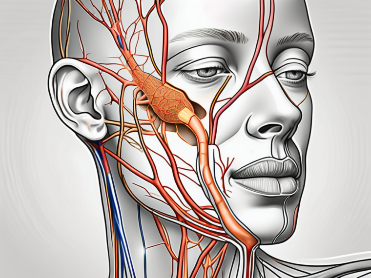
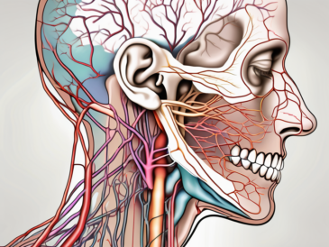
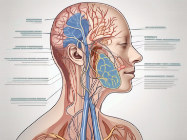
The glossopharyngeal nerve, a crucial component of the human nervous system, serves a vital role in innervating various structures within the head and neck. Among its many responsibilities, one of its notable functions is the innervation of the parotid gland, a significant salivary gland situated in close proximity to the ear.
Understanding the Glossopharyngeal Nerve
The glossopharyngeal nerve, designated as cranial nerve IX, emerges from the medulla oblongata, descending through the jugular foramen, and eventually branching out to innervate specific regions of the head and neck. Its intricate anatomy and diverse functions make it a fascinating subject of study for medical professionals.
The glossopharyngeal nerve is a crucial component of the cranial nerves, a set of twelve nerves that emerge directly from the brain. It is one of the four cranial nerves involved in the control of the muscles responsible for swallowing and speech production. These motor fibers of the glossopharyngeal nerve innervate the stylopharyngeus muscle, which plays a vital role in the swallowing process.
But the glossopharyngeal nerve is not just limited to motor functions. It also contains sensory fibers that transmit information from various regions of the head and neck to the brain. These sensory fibers are responsible for relaying taste sensations from the posterior third of the tongue, making the glossopharyngeal nerve an essential player in the perception of taste.
Anatomy of the Glossopharyngeal Nerve
The glossopharyngeal nerve comprises motor, sensory, and autonomic fibers. Motor fibers primarily innervate muscles involved in swallowing and speech production. Sensory fibers transmit information from the tongue, tonsils, pharynx, and middle ear to the brain. Autonomic fibers regulate parasympathetic functions, controlling salivation, blood pressure, and heart rate.
The motor fibers of the glossopharyngeal nerve originate from the nucleus ambiguus, a cluster of nerve cell bodies located in the medulla oblongata. From there, they travel through the glossopharyngeal nerve and eventually reach the stylopharyngeus muscle, which is responsible for elevating the pharynx during swallowing.
The sensory fibers of the glossopharyngeal nerve have a wide distribution, covering various regions of the head and neck. They receive sensory input from the posterior third of the tongue, tonsils, pharynx, and middle ear. This information is then transmitted to the brain, allowing for the perception of taste, touch, and sound.
Additionally, the glossopharyngeal nerve also contains autonomic fibers that regulate parasympathetic functions. These fibers control salivation, ensuring the proper lubrication of the oral cavity during the digestive process. They also play a role in regulating blood pressure and heart rate, contributing to the overall cardiovascular health of an individual.
Functions of the Glossopharyngeal Nerve
Aside from its role in innervating the parotid gland, the glossopharyngeal nerve also contributes to taste perception on the posterior third of the tongue, monitors blood pressure and oxygen levels, and facilitates the gag reflex. Its multifaceted functions highlight its significance within the human body.
The glossopharyngeal nerve plays a crucial role in the perception of taste. The sensory fibers of the nerve receive taste information from the posterior third of the tongue, allowing individuals to experience the different flavors of food and beverages. This ability to taste is not only essential for enjoyment but also for detecting potential dangers, such as spoiled or toxic substances.
In addition to taste perception, the glossopharyngeal nerve also monitors blood pressure and oxygen levels within the body. It contains specialized receptors called baroreceptors and chemoreceptors, which constantly assess the state of these vital parameters. If there is a significant deviation from the normal range, the glossopharyngeal nerve sends signals to the brain, triggering appropriate responses to maintain homeostasis.
Furthermore, the glossopharyngeal nerve is involved in the gag reflex, a protective mechanism that helps prevent choking. When an object or substance touches the back of the throat, the sensory fibers of the glossopharyngeal nerve detect the stimulus and initiate a reflexive contraction of the muscles in the throat. This reflexive action helps expel the foreign object or substance, ensuring the airway remains clear.
The glossopharyngeal nerve’s diverse functions highlight its importance in various physiological processes. From taste perception to blood pressure regulation and the gag reflex, this cranial nerve plays a significant role in maintaining the overall health and well-being of an individual.
The Parotid Gland: An Overview
The parotid gland, the largest of the salivary glands, sits just in front of the ear, extending towards the lower jaw. It plays a crucial role in producing saliva, which aids in digestion and maintains oral health. Understanding the location, structure, and functions of the parotid gland provides valuable insights into its interplay with the glossopharyngeal nerve.
Location and Structure of the Parotid Gland
The parotid gland is nestled within the parotid space, which encompasses the area between the skin, the mastoid process, and the mandible. This space provides a protective environment for the gland, shielding it from external factors that may cause damage or infection.
The parotid gland itself is composed of serous acinar cells, which are responsible for producing saliva. These cells are arranged in lobules, forming the distinct structure of the gland. The lobules are connected by connective tissue, providing support and maintaining the overall integrity of the gland.
Within the parotid gland, there are numerous ducts that transport saliva from the acinar cells to the oral cavity. The Stensen’s duct, also known as the parotid duct, is the main duct responsible for carrying saliva. It begins at the gland and travels through the cheek, opening into the oral cavity near the second upper molar.
Role and Functions of the Parotid Gland
The primary function of the parotid gland is to secrete saliva, facilitating the initial stages of food digestion, lubrication, and neutralization of oral pH. Saliva is essential for the breakdown of food particles, making them easier to swallow and digest. It also helps in the formation of the bolus, the mass of food that is formed in the mouth and swallowed.
Saliva produced by the parotid gland contains various enzymes, including amylase, which aids in the breakdown of complex carbohydrates. This enzymatic action begins the process of carbohydrate digestion even before the food reaches the stomach. The parotid gland’s contribution to efficient nutrient absorption cannot be overstated.
In addition to its role in digestion, the parotid gland also plays a vital role in maintaining oral health. Saliva helps to cleanse the oral cavity, washing away food debris and bacteria that may lead to tooth decay and gum disease. It also contains antimicrobial properties, further protecting the mouth from harmful microorganisms.
Furthermore, the parotid gland is involved in temperature regulation within the oral cavity. The production of saliva helps to cool down the mouth, especially during periods of increased heat or physical exertion. This cooling effect provides comfort and prevents overheating of the oral tissues.
The interplay between the parotid gland and the glossopharyngeal nerve is crucial for the regulation of saliva production. The glossopharyngeal nerve, one of the cranial nerves, carries sensory information from the parotid gland to the brain. This feedback loop allows the brain to monitor and adjust saliva production based on the body’s needs.
In conclusion, the parotid gland is a remarkable organ that serves multiple functions in the body. Its location, structure, and functions all contribute to the overall well-being of the oral cavity and the digestive system as a whole. Understanding the intricate details of the parotid gland enhances our appreciation for its essential role in maintaining oral health and facilitating efficient digestion.
Innervation of the Parotid Gland by the Glossopharyngeal Nerve
The innervation of the parotid gland by the glossopharyngeal nerve is a crucial aspect of its proper functioning. The intimate connection between these two structures highlights the significance of coordinated neural signaling.
The parotid gland, located in front of the ear and extending into the cheek, is the largest of the salivary glands. It plays a vital role in the production and secretion of saliva, which is essential for the initial digestion of food and maintaining oral health. The glossopharyngeal nerve, one of the cranial nerves, is responsible for providing the necessary innervation to the parotid gland.
Process of Innervation
The glossopharyngeal nerve carries parasympathetic fibers that synapse with the postganglionic parotid plexus within the gland. This intricate network of nerve fibers allows for precise control over the secretion of saliva. When stimulated, the parasympathetic fibers trigger the serous acinar cells of the parotid gland to produce and release saliva.
The parotid plexus, a complex network of nerves, is responsible for transmitting the signals from the glossopharyngeal nerve to the various parts of the parotid gland. It ensures that the saliva production is regulated and coordinated, allowing for optimal functioning of the gland.
Saliva, composed of water, electrolytes, enzymes, and antibacterial substances, not only aids in the initial digestion of food but also helps in maintaining oral hygiene. It lubricates the oral cavity, facilitating speech and swallowing, and protects the teeth and oral mucosa from harmful bacteria.
Importance of the Glossopharyngeal Nerve in Parotid Gland Function
The glossopharyngeal nerve plays a vital role in regulating salivary production. Dysfunction or damage to the nerve can lead to diminished saliva production, resulting in dry mouth (xerostomia), difficulty in swallowing, and potentially oral health issues.
Individuals with xerostomia may experience discomfort, difficulty in speaking, altered taste sensation, and an increased risk of dental caries and oral infections. The lack of saliva can also affect the overall health of the oral cavity, as it is an important defense mechanism against harmful microorganisms.
Medical professionals should promptly address any impairments or damage to the glossopharyngeal nerve to prevent further complications. Treatment options may include medications to stimulate saliva production, lifestyle modifications, and addressing the underlying cause of the nerve dysfunction.
In conclusion, the innervation of the parotid gland by the glossopharyngeal nerve is a complex and essential process for the proper functioning of the gland. The precise coordination between the nerve fibers and the parotid plexus ensures the regulated production and release of saliva, which is crucial for maintaining oral health and overall well-being.
Disorders Related to the Glossopharyngeal Nerve and Parotid Gland
While the interplay between the glossopharyngeal nerve and the parotid gland is usually harmonious, certain disorders can disrupt their normal functioning. Prompt recognition, diagnosis, and management are essential in ensuring optimal health outcomes.
The glossopharyngeal nerve, also known as the ninth cranial nerve, plays a crucial role in various functions related to the throat and tongue. It is responsible for transmitting sensory information from the back of the throat, including taste, to the brain. Additionally, it helps control the muscles involved in swallowing and speech.
The parotid gland, on the other hand, is the largest of the salivary glands and is located in front of the ear. It produces saliva, which aids in the digestion of food and helps maintain oral health. The glossopharyngeal nerve innervates the parotid gland, allowing it to function properly.
Symptoms and Diagnosis
Signs of glossopharyngeal nerve or parotid gland disorders may include difficulty swallowing, dry mouth, ear pain, or even a visible swelling around the jaw area. These symptoms can significantly impact an individual’s quality of life and may require medical attention.
When a patient presents with these symptoms, a comprehensive medical evaluation is necessary to accurately diagnose the underlying condition. The healthcare provider will conduct a thorough physical examination, assessing the patient’s ability to swallow, checking for any visible abnormalities, and evaluating the function of the parotid gland.
In some cases, imaging studies such as an MRI or CT scan may be ordered to obtain detailed images of the glossopharyngeal nerve and parotid gland. These imaging techniques can help identify any structural abnormalities or lesions that may be causing the symptoms.
Furthermore, additional tests, such as a swallow study or a biopsy of the parotid gland, may be performed to gather more information about the specific disorder affecting the glossopharyngeal nerve or parotid gland.
Treatment and Management
The treatment approach for glossopharyngeal nerve or parotid gland disorders depends on the specific condition diagnosed. It is essential to consult with a qualified healthcare professional who can provide personalized advice and tailored treatment plans.
In some cases, medication may be prescribed to alleviate symptoms and manage the underlying condition. For example, if the disorder is causing dry mouth, medications that stimulate saliva production may be recommended. Pain medications or anti-inflammatory drugs may be prescribed to relieve ear pain or reduce swelling.
Physical therapy can also play a significant role in the management of glossopharyngeal nerve or parotid gland disorders. Therapeutic exercises can help improve swallowing function and strengthen the muscles involved in speech production.
Lifestyle modifications may be suggested to minimize symptoms and promote overall well-being. These may include dietary changes, such as consuming softer foods or avoiding certain triggers that exacerbate symptoms. Maintaining good oral hygiene and staying hydrated can also be beneficial.
In more severe cases, surgical interventions may be necessary. Surgery can be performed to remove tumors or correct structural abnormalities that are causing the glossopharyngeal nerve or parotid gland dysfunction. The specific surgical procedure will depend on the individual’s condition and needs.
Regular follow-up appointments with the healthcare provider are essential to monitor the progress of treatment and make any necessary adjustments. Open communication between the patient and the healthcare team is crucial to ensure optimal management of glossopharyngeal nerve or parotid gland disorders.
Conclusion: The Interplay between the Glossopharyngeal Nerve and the Parotid Gland
The glossopharyngeal nerve and the parotid gland share a fascinating relationship within the human body. With the glossopharyngeal nerve responsible for innervating the parotid gland and maintaining its proper functioning, any disturbances in this intricate interplay can have significant implications for overall health. Therefore, it is essential to recognize the signs of any related disorders and seek medical advice promptly. By doing so, individuals can ensure the well-being of their glossopharyngeal nerve and parotid gland, promoting optimal oral health and overall quality of life.
